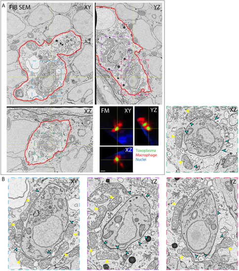
Host cell-intrinsic response disrupts Toxoplasma gondii parasitophorous vacuoles in zebrafish macrophages in vivo. (A) 3D CLEM of two parasites inside a macrophage in the HBV of mpeg1:G/U:mCherry larvae harboring macrophages (red) infected with type I Toxoplasma-GFP (green) at 6 hpi and stained with Hoechst 33342 (blue). Orthoslices of 3200 consecutive 5 nm FIB SEM slices of the whole macrophage (top left; see also Movie 7) and of 59 confocal z-slices (40×) re-sliced to match the orientation of the FIB SEM data (FM, bottom right; cell also shown in yellow dashed line box in Fig. 6A). Color boxes show localization of the cropped and enlarged images of tachyzoites shown in B. The plasma membrane of the macrophage (red) was manually segmented to aid correlation. (B) Cropped and enlarged images of the Toxoplasma shown in the FIB SEM orthoslices in A. Host mitochondrial recruitment to the PV is indicated by yellow arrowheads; breaks in the PV are indicated by cyan arrowheads. Scale bars: 5 µm (A) and 1 µm (B).
|
