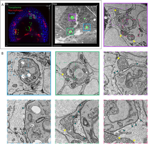
Host cell-intrinsic response in zebrafish disrupts Toxoplasma gondii parasitophorous vacuoles in brain cells in vivo. (A) 3D CLEM of tachyzoites in the HBV of transgenic mpeg1:G/U:mCherry larvae harboring macrophages (red) infected with type I Toxoplasma-GFP (green) at 6 hpi and stained with Hoechst 33342 (blue). 3D reconstructions of 59 confocal z-slices (40×) of a part of the HBV (FM, fluorescence microscopy; left) and of 75 inverted consecutive 70 nm sections imaged by ssTEM at 440× magnification (right). Each of the Toxoplasma visible in the ssTEM dataset was manually segmented (green; right) in every section to aid correlation. Regions of interest showing the localization of the high-resolution ssTEM images shown in B (cyan, magenta and green boxes) and Fig. 7 (yellow dashed line box) are indicated. Scale bars: 10 µm. (B) Representative higher-magnification ssTEM images of Toxoplasma in HBV cells. Continuous line boxes show 6800× magnification images; dashed line boxes show 18,500× magnification images. Toxoplasma tachyzoites were imaged in their full volume to accurately assess the continuity of the PV. See also Movie 6. Host mitochondrial recruitment to the PV is indicated by yellow arrowheads; breaks in the PV are indicated by cyan arrowheads. Scale bars: 1 µm (continuous line box) and 500 nm (dashed line box).
|
