|
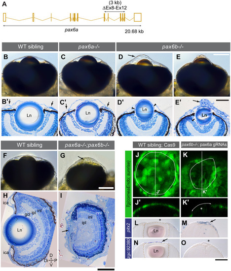
Pax6a/b dose correlates with ASD severity.(A) CRISPR/Cas9-mediated deletion of 3 kb in the pax6a locus encompassing exons 8–12 (ΔEx8-Ex12, a horizontal double head arrow) encoding homeobox DNA binding and PST domains (S11 Fig). (B-E’) The AS phenotype at 5 dpf of a wild type sibling (B-B’), a pax6a-/- zygotic mutant (C-C’) and pax6b-/- maternal zygotic mutants with keratoconus-like phenotype (D-D’; arrow in D) and deflated cornea (E-E’), imaged alive (B-E) and in sections (B’-E’). (B’-E’) The chamber angle (arrows in B’-C’) is open in wild type (B’), pax6a-/- (C’) and pax6b-/- embryos with keratoconus (D’), however, it appears closed in pax6b-/- embryos with deflated cornea (E’). Although a pax6a-/- embryo appeared normal with live inspection (C), in comparison to the wild type that has a flat iris plane (B’) the iris plane is slanted (C’), as also seen in pax6b-/- embryos (D’-E’). All pax6 mutants show a smaller lens (Ln). (D’-E’) Gaps are evident between the lens capsule and the lens fibre (arrowheads). Abnormal layering in the corneal epithelium is occasionally observed with deflated corneas (arrow in E’). (F-I) The AS phenotype at 5 dpf of a wild type sibling (F, H) and a double homozygous pax6a-/-;pax6b-/- embryo (G, I). ica: iridocorneal angle, gcl: ganglion cell layer, ipl: inner plexiform layer, inl: inner nuclear layer, onl: outer nuclear layer. The distal eye structure of pax6a/b double mutants has no lens, no gcl, no ica and is occupied with abnormal ocular mesenchymal cells (arrows in G, I). Orientation is dorsal (D) up, ventral (V) down, distal (Di) left and proximal (P) right. (J-O) Marker gene expression analysis in 5 dpf embryos of wild type siblings injected with Cas9 without gRNAs (J, J’, L, and N) and of maternal zygotic pax6b-/- mutant genotype injected with pax6a gRNAs and Cas9 (K, K’, M and O). (J-K’) The corneal endothelium visualized by the Tg(corneaEndo:GFP) transgenic line. The anterior chamber margin is demarcated by a solid circle. Optical sections made along vertical lines in J and K are shown in J’ and K’, respectively. The Pax6 deficient embryo (K-K’) lacked both the lens (asterisk in K’) and the expression of the corneal endothelium reporter (K’, double arrowed arc). (L-O) in situ expression analysis of a mesenchymal marker pitx2 (L-M) and of the corneal epithelium marker zgc:92380 (N-O). Presence and absence of gene expression is denoted by arrow and asterisk, respectively. The lens border is demarcated by a stippled line. Scale bars: 50 μm.
|