|
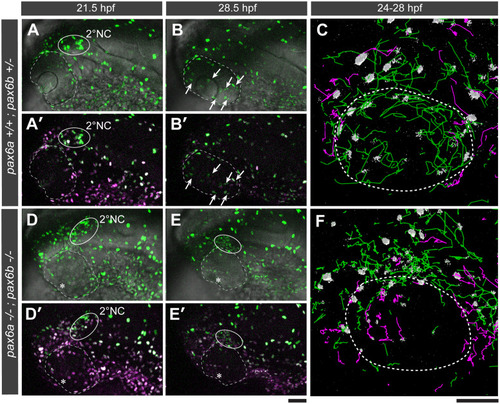
Pax6a/b are required for guiding the two ocular NC populations to their distinct destinations.Tg(sox10:h2a-tdEosFP) embryos with a genetic background of either pax6a+/+;pax6b+/- (A-C, wild type-like) or pax6a-/-;pax6b-/- (D-F, double homozygous mutant) were photoconverted at 17 hpf for visualizing 1° and 2° NC cells in magenta and green, respectively. At 21.5 hpf, the 2°NC cell cluster (A-A’ and D-D’; solid circles) is observed at the dorsal edge of the eye in both genotypes. At 28.5 hpf, although 2°NC cluster was dissolved into individually migrating 2°NC cells in the distal eye compartment of a pax6a+/+;pax6b+/- embryo (B-B’, arrows), the 2°NC cluster persisted at the same location in a pax6a-/-;pax6b-/- mutant embryo (E-E’, solid circle) which failed to develop the lens primordium (D-E’, asterisks). (C and F) Lateral views of 3D rendered trajectories of 1°NC (magenta lines) and 2°NC cells (green lines) for each genotype during 24–28 hpf. The NC nucleus is represented as a grey object. The eye boundary is shown as a stippled circle. Scale bars: 50 μm.
|