
Fig. 12
- ID
- ZDB-FIG-200327-24
- Publication
- Li et al., 2019 - Development and organization of the zebrafish intestinal epithelial stem cell niche
- Other Figures
- All Figure Page
- Back to All Figure Page
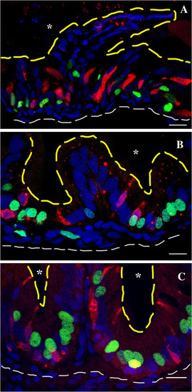
Secretory cells interdigitate between proliferating epithelial cells between 20 and 33 dpf. Labeling with Edu on three different days during the third week postembryogenesis labels multiple cells at the interfold base (A). Secretory cells now interdigitate between many of the proliferating epithelial cells. At 33 dpf, a single pulse of Edu labels multiple cells at the interfold base (B). Similar to 26 dpf, the interfold base now has many secretory cells and proliferating epithelial cells interdigitated. Numerous interdigitating secretory cells are observed between proliferating epithelial cells in the adult intestine following a single Edu pulse (C). The edge of the fold facing the lumen is outlined with the yellow dashed line. Red, pan‐secretory 2F11; green, Edu; blue, DAPI; white dashed line outer edge of intestine, asterisk, intestinal lumen. Scale bar = 10 μm. |