
FIGURE
Fig. 7
- ID
- ZDB-FIG-200327-19
- Publication
- Li et al., 2019 - Development and organization of the zebrafish intestinal epithelial stem cell niche
- Other Figures
- All Figure Page
- Back to All Figure Page
Fig. 7
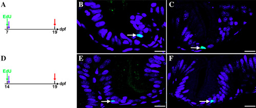
Position of proliferating cells between 6 and 19 dpf. Epithelial cells are labeled with a pulse of Edu (1 hr) at 7 dpf (A, blue arrow) and 14 dpf (D, blue arrow) followed by a chase to 19 dpf (red arrow). All labeled cells start in the interfold base (B, 7dpf and E, 14 dpf) and remain at this position following the chase period (C, 7 to 19 dpf and F, 14 to 19 dpf). Arrows indicate Edu‐labeled cells (B, C, E, and F); Scale bar = 10 μm; n = 5. |
Expression Data
Expression Detail
Antibody Labeling
Phenotype Data
Phenotype Detail
Acknowledgments
This image is the copyrighted work of the attributed author or publisher, and
ZFIN has permission only to display this image to its users.
Additional permissions should be obtained from the applicable author or publisher of the image.
Full text @ Dev. Dyn.