
Fig. 9
- ID
- ZDB-FIG-200327-21
- Publication
- Li et al., 2019 - Development and organization of the zebrafish intestinal epithelial stem cell niche
- Other Figures
- All Figure Page
- Back to All Figure Page
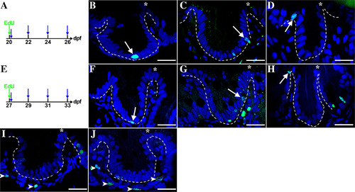
A–J: Epithelial cells migrate up the folds during the third and the fourth week postembryogenesis. Individuals are injected with Edu (green arrow) either at 20 (A) or 27 (E) dpf. Samples are taken at 1 hr to determine initial cell position (first blue arrow, A and E) and then at 2‐day intervals to the end of the week (three additional blue arrows, A and E). With a pulse of Edu at either 20 dpf (A) or 27 dpf (D) epithelial cells are labeled only at the fold base (arrows). Following a chase for 2 days to either 22 (C) or 29 (G) dpf, progeny reach the mid‐fold (arrows) and reach the tip by 4 days at either24 (B) or 31 (E) dpf. In each of the histological panels, the dashed line is drawn between the epithelial and mesenchymal layers. During these 2 weeks, mesenchymal cells are also labeled (examples at 24 dpf‐C and 31‐F; mesenchymal cells indicated with arrowheads); n = 4. |