
Fig. 1
- ID
- ZDB-FIG-200327-16
- Publication
- Li et al., 2019 - Development and organization of the zebrafish intestinal epithelial stem cell niche
- Other Figures
- All Figure Page
- Back to All Figure Page
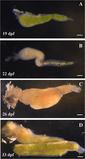
Growth of postembryonic larval intestine. The larval intestine continues as a straight tube for the 2 weeks following embryogenesis (A). The main change in appearance of the intestine between 6 and 19 dpf is doubling or tripling of the intestinal diameter. During the third week (20 to 26 dpf), a loop develops by first bending the intestine laterally and then rotating the bend dorsally (B). The loop continues to grow anteriorly and appears to draw in a portion of the posterior intestine into the loop (C). The intestine continues to grow in length during the fourth week (27 to 33 dpf) but not at the same rate as in the third week. One of the changes is a narrowing of the anterior intestinal diameter (D). Scale bar = 100 μm |