
Fig. 11
- ID
- ZDB-FIG-200327-23
- Publication
- Li et al., 2019 - Development and organization of the zebrafish intestinal epithelial stem cell niche
- Other Figures
- All Figure Page
- Back to All Figure Page
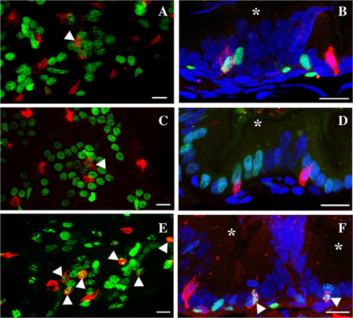
Secretory cells associate with clusters of proliferating intestinal epithelial cells. A pulse of Edu at 96 hpf followed by a chase to 120 hpf labels a group of proliferating cells (A) which is present at the fold base (B). Secretory cells (anti‐2F11) are interspersed within proliferating epithelial cells. Labeling with Edu on three different days during the first week postembryogenesis (C) (6–12 dpf) or the second week postembryogenesis (E) (13–19 dpf) labels clusters of proliferating epithelial cells. Secretory cells develop within groups of proliferating cells at the fold base (D) (6–12 dpf) and (F) (13–19 dpf) in a similar manner and number as observed at the end of embryogenesis. Some fold base epithelial cells begin differentiating into secretory cells (arrowhead A, C, E, and F) as they co‐label with 2F11 and Edu. Pan‐secretory 2F11, red; Edu, green; DAPI, blue; asterisk, intestinal lumen. Anterior is to the left and dorsal to the top in A, C, and E. Scale bar = 10 μm. |