
Fig. 4
- ID
- ZDB-FIG-200325-195
- Publication
- Bagwell et al., 2020 - Notochord vacuoles absorb compressive bone growth during zebrafish spine formation
- Other Figures
- All Figure Page
- Back to All Figure Page
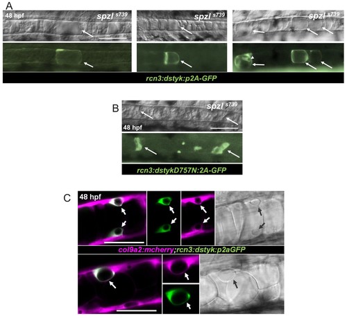
Dstyk’s kinase activity is required cell-autonomously for vacuole formation.( A) Live DIC image (top) and live fluorescent image (bottom) of spzl mutants injected with a construct driving dstyk:p2aGFP expression in the notochord. The arrows point to GFP expressing cells with rescued vacuole integrity. Arrow head points to an expressing sheath cell that also develops a vacuole. WT dstyk rescued vacuole integrity in 90% of expressing cells (n = 30). Scale bar = 100 µm. ( B) Live DIC image (top) and fluorescent image (bottom) of a spzl-/- embryo injected with dstykD757N:p2aGFP, a construct driving a (D757N) kinase dead version of dstyk in the notochord. Expressing cells (arrows) exhibit fragmented vacuoles and only 5% rescue (n = 20). Scale bar = 100 µm. ( C) Confocal and DIC images of WT embryos at 48 hpf expressing col9a2:mcherry in the notochord sheath and injected with dstyk:p2aGFP. Sheath cells expressing dstyk:p2aGFPdevelop a vacuole. Arrows point to the vacuole. Scale bar = 50 µm. |