
Fig. 1
- ID
- ZDB-FIG-200325-191
- Publication
- Bagwell et al., 2020 - Notochord vacuoles absorb compressive bone growth during zebrafish spine formation
- Other Figures
- All Figure Page
- Back to All Figure Page
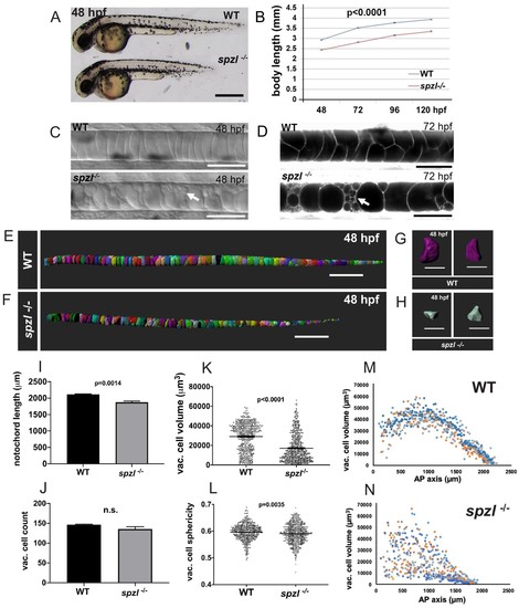
spzl is a recessive mutation which causes notochord vacuole fragmentation, impaired axis elongation, and altered vacuolated cell packing.( A) Whole mount lateral view of 48 hpf spzl-/- (bottom) and WT sibling (top) embryos. Scale bar = 500 µm. ( B) Body length measurements (mm) from 48 to 120 hpf. n = 30 for WT and n = 27, n = 30, n = 29, n = 28 for spzl-/- respectively. p<0.0001 at all time points, two-way ANOVA with Sidak’s test. At 24 hpf spzl mutant embryos (n = 20) are also significantly shorter than WT (n = 15), p=0.001, unpaired t-test using Welch’s correction. ( C) Live DIC images of 48 hpf WT (top) and spzl-/- (bottom) embryos. Arrow points to fragmented vacuoles. Scale bars = 50 µm. ( D) Live confocal images of 72 hpf WT (top) and spzl-/- (bottom) notochords stained with Cell Trace to visualize internal membranes. Arrow points to area of vacuole fragmentation. Scale bars = 50 µm. ( E–F) Notochord 3D reconstructions for 48 hpf WT ( E) and spzl-/- ( F) embryos. Scale bar = 200 µm. ( G–H) Single cell 3D reconstructions for WT ( G) and spzl-/- ( H) visualized at different angles to show cell shape. Scale bar = 50 µm. ( I) Notochord length measurements for WT and spzl-/- at 48 hpf. ( J) Total number of vacuolated cells in WT and spzl-/- at 48 hpf. ( K) Plot of cell volume measurements of WT and spzl-/-notochord cells at 48 hpf. ( L) Sphericity of individual notochord cells for WT and spzl-/- at 48 hpf. ( M–N) Volume of notochord cells along the AP axis of WT and spzl-/- at 48 hpf. p-values were determined by an un-paired t-test using Welch’s correction. |
| Fish: | |
|---|---|
| Observed In: | |
| Stage Range: | Long-pec to Day 5 |