FIGURE

Figure 5

ID
ZDB-FIG-200318-14
Publication
Weinberger et al., 2020 - Functional Heterogeneity within the Developing Zebrafish Epicardium
Other Figures
All Figure Page
Back to All Figure Page
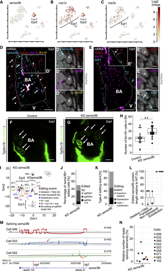
Figure 5

Semaphorin 3fb Is a Marker of Epi2 and Controls the Number of tbx18+ Cells in the Bulbus Arteriosus

(A–C) Expression of sema3fb (A), nrp1a (B), and nrp2a (C). Color key indicates range of log2 transformed FPKM values.

(D) mRNA staining of sema3fb (cyan), nrp2a (red, arrowheads), nrp1a (magenta, arrows), and tbx18 (orange) at 5 dpf. (D′) Close proximity of nrp2a, nrp1a, and tbx18 to a nucleus (asterisk) at the BA boundary.

(E) mRNA staining of nrp2a (red), nrp1a (magenta), and tcf21 (cyan) at 5 dpf. (E′) A nucleus (asterisk) in close proximity to nrp2a, nrp1a, and tcf21.

(F) The BA in a 5 dpf control larva.

(G) Increased numbers of tbx18:myr-Citrine+ cells (arrows) in the BA of a KO sema3fb larva.

(H) Absolute quantification of tbx18:myr-Citrine+ cell numbers in the BA at 5 dpf.

(I–N) Analysis of gene editing in single sema3fb-mutated epicardial cells. (I) t-SNE plot showing cell clusters and editing events. Numbers indicate cell identities, and colors depict editing events. Ld, large deletion; fs, frameshift; and sub, substitution. (J) Relative number of Cas9-edited sema3fb+ cells. Colors indicate the sema3fb sgRNA target site edited. (K) Frequency of editing event types. (L) Inferred edited sema3fb protein lengths, relative to WT. (M) Sashimi plots showing splicing of sema3fb. Arrows highlight reads splicing abnormally. Red bars indicate sgRNA target sites. (N) Relative number of sema3fb splicing reads that spliced abnormally.

Scale bars: 10 μm in (D)–(G) and 5 μm in (D′) and (E′). Color channels were adjusted separately for brightness and contrast. (D)–(G) are single optical sections. Data in (H) and (L) are represented as median, first, and third quartiles (box). Significance calculated using Welch’s t test. ∗∗p < 0.01. V, ventricle; BA, bulbus arteriosus.

See also Figure S6.

Expression Data
Genes:
Fish:
Knockdown Reagents:
Anatomical Term:
Stage: Day 5

Expression Detail
Antibody Labeling
Phenotype Data
Fish:
Knockdown Reagents:
Observed In:
Stage: Day 5

Phenotype Detail
Acknowledgments
This image is the copyrighted work of the attributed author or publisher, and ZFIN has permission only to display this image to its users. Additional permissions should be obtained from the applicable author or publisher of the image.

Reprinted from Developmental Cell, 52(5), Weinberger, M., Simões, F.C., Patient, R., Sauka-Spengler, T., Riley, P.R., Functional Heterogeneity within the Developing Zebrafish Epicardium, 574-590.e6, Copyright (2020) with permission from Elsevier. Full text @ Dev. Cell