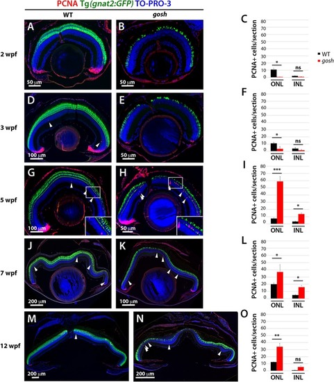
Proliferation of Müller glia, NPCs, and rod precursors starts after 5 wpf in gosh mutants. Labeling of wild-type sibling (A,D,G,J,M) and gosh mutant retinas (B,E,H,K,N) at 2, 3, 5, 7, and 12 wpf with anti-PCNA antibody. Cones are visualized with the Tg(gnat2:GFP) transgenic line, and nuclei are counterstained with TO-PRO-3. In wild-type retinas, retinal stem and progenitor cells in the CMZ express PCNA, indicating proliferation of retinal progenitor cells. Furthermore, PCNA-positive cells are observed in the ONL and INL, corresponding to rod precursors, and Müller glia/NPCs, respectively, at all stages studied (A,D,G,J,M). In gosh mutants, PCNA expression is absent in the CMZ, as well as in the ONL and INL at 2–3 wpf (B,E). At 5 wpf, PCNA-positive ONL and INL cells are drastically increased in gosh mutants, suggesting that Müller glia and rod precursor cells start cell proliferation (H). Insets show PCNA-positive cells (G,H). Numbers of PCNA-positive cells in the ONL and INL indicate that proliferation becomes maximal at 5 wpf in gosh mutants and maintains higher levels than wild type until 12 wpf (C,F,I,L,O). Bars and lines indicate mean ± SEM, n: 3–9. Black and red bars: wild-type sibling and gosh mutant. Arrowhead depict PCNA-positive cells in the INL (ns, p > 0.05; ∗p < 0.05; ∗∗p < 0.01; and ∗∗∗p < 0.001).
|
