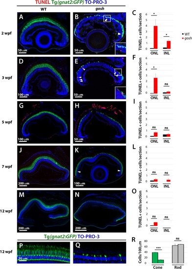
Retinal apoptosis occurs transiently at 2–3 wpf and ceases by 5 wpf in gosh mutants. TUNEL was assessed on retinal slides of wild-type sibling (A,D,G,J,M) and gosh mutant retinas (B,E,H,K,N) at 2, 3, 5, 7, and 12 wpf. Apoptotic cells in the ONL are indicated by arrows. The transgenic line Tg(gnat2:GFP) is used to label cone photoreceptors. Nuclei are counterstained with TO-PRO-3. Wild-type sibling retinas show very low numbers of TUNEL-labeled cells at all stages (A,D,G,J,M). In gosh mutants, numbers of apoptotic cells are higher in the ONL at 2–3 wpf and in the INL at 2 wpf (B,E, insets show TUNEL+ cells in the ONL). Quantification of TUNEL-positive nuclei in the ONL and INL was performed (C,F,I,L,O). Bars and lines indicate means ± SEM, n: 3–7. Black and red bars: wild-type sibling and gosh mutants. Central retina of control and gosh retinas at 12 wpf show similar rod layer thickness, but cone photoreceptor shows a reduce number of cones (P–R, control left bars, gosh right bars) (ns, p > 0.05; ∗p < 0.05; and ∗∗∗p < 0.001). ONL, outer nuclear layer; INL, inner nuclear layer.
|
