
Fig. 2
- ID
- ZDB-IMAGE-200201-13
- Antibodies
- Publication
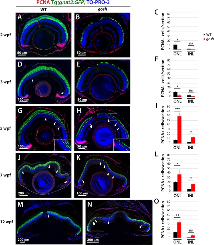
- Iribarne et al., 2019 - TNFα Induces Müller Glia to Transition From Non-proliferative Gliosis to a Regenerative Response in Mutant Zebrafish Presenting Chronic Photoreceptor Degeneration
- All Figures
- Figures for Iribarne et al., 2019
|
Fig. 2
Proliferation of Müller glia, NPCs, and rod precursors starts after 5 wpf in