|
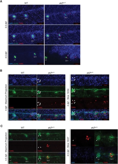
Increased cell death in the otic vesicles and the PLL of ak2hg15 embryos. (A) Maximum projections of 3.5 and 4.5 dpf ak2hg15 embryos and their control siblings in the Tg(pou4f3:GAP-GFP) background (indicated as pou4f3) stained with TUNEL assays (red signal). (B,C) Maximum projections (left panels) and representative single plane confocal analysis (right panels) at 4 (B) and 4.5 (C) dpf of a TUNEL assay (red signal) performed on ak2hg15 embryos and their siblings in the Tg(-8.0cldnb:LY-EGFP) background (cldnb, green) labeling the migrating secondary primordium (dashed ellipse), deposited primary and secondary (red circle) neuromasts and epithelial cells. White arrowheads label TUNEL-positive cells in the L1 primary neuromast. Nuclei are labeled with DAPI (blue). Scale bars: 50 µm (A,B); 20 µm (C).
|