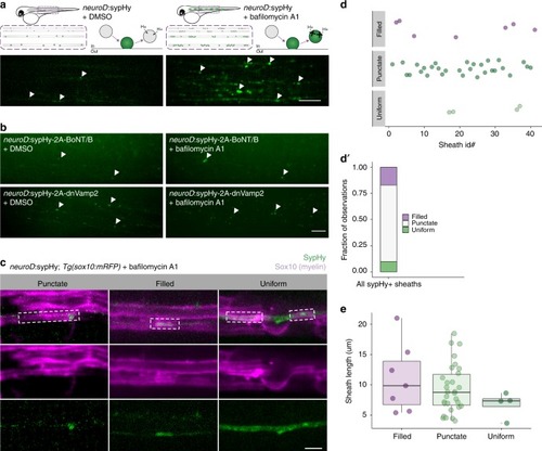
Variable synaptic vesicle exocytosis sites under myelin sheaths. a Left, schematic of Syp-pHluorin (SypHy) function and transient expression in spinal cord neurons and representative max-projection image of a live 4 dpf larva expressing neuroD:sypHy. Arrowheads indicate dim spots of SypHy signal. Right, similar but in the presence of bafilomycin A1 to inhibit vesicle reacidification. Note appearance of several large SypHy + “hotspots” along axons. Scale bar, 10 µm. b Similar to a except larvae are co-expressing inhibitors of exocytosis, botulinum toxin (BoNT/B) or dominant-negative Vamp2 (dnVamp2). Both BoNT/B and dnVamp2 suppress bafilomycin induction of SypHy hotspots. Arrowheads indicate a few visible SypHy spots. Scale bar, 10 µm. c Expression of neuroD:sypHy in Tg(sox10:mRFP) larvae treated with bafilomycin reveals three types of SypHy-reported exocytosis sites under myelin sheaths: punctate, filled, and uniform. Boxes outline individual myelin sheaths. Scale bar, 5 µm. d Classification plot of SypHy signal under 41 sheaths (from n = 20 larvae) by category. Presented as a bar in d′ for visible contribution of each category to the total. e No association between category and sheath length, Kruskal–Wallis test
|
