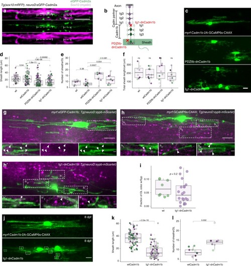
The extracellular, trans-acting adhesion domain (Ig1) of Cadm1b promotes myelin sheath growth. a Neuronal eGFP-Cadm2a puncta (green) localize under myelin sheaths (magenta, brackets). Scale bar, 10 µm. b Schematic of dominant-negative oligodendrocyte Cadm1b variants, PDZIIb-dnCadm1b and Ig1-dnCadm1b, interacting with partner Cadm located on neurons. c Representative examples of oligodendrocytes expressing wildtype Cadm1b, PDZIIb-dnCadm1b, and Ig1-dnCadm1b with GCaMP6s-CAAX to label sheath membrane. Scale bar, 10 µm. d Sheath lengths for wt, wtCadm1b-, PDZIIb-dnCadm1b-, and Ig1-dnCadm1b-expressing oligodendrocytes. Note that PDZIIb-dnCadm1b is the same allele presented in Fig. 4. n (cells/sheaths) = 8/74 (wt), 11/91 (wtCadm1b), 10/159 (PDZIIb-dnCadm1b), 10/115 (Ig1-dnCadm1b), Wilcox rank-sum test with Bonferroni-Holm correction for multiple comparisons. Dots of the same color indicate sheaths belonging to the same cell and match those in plots e and f. e Sheath number for wt, wtCadm1b-, PDZIIb-dnCadm1b-, and Ig1-dnCadm1b-expressing oligodendrocytes. Same n and statistical test as in d. f Total sheath length generated per cell is unchanged by all alleles, Kruskal–Wallis test. (g, h, h′) Max projection image of myrf:eGFP-Cadm1b oligodendrocyte in Tg(neuroD:sypb-mScarlet) larvae, in which Syp-mScarlet is expressed pan-neuronally. Inset is a substack projection of 6 slices (0.31 µm/slice) containing eGFP-Cadm1b-labeled sheaths wrapping Syp-mScarlet puncta. Arrowheads indicate colocalized oligodendrocyte eGFP-Cadm1b and neuronal Syp-mScarlet signal. h, h′ myrf:GCaMP6s-CAAX (wt) (h) or Ig1-dnCadm1b (h′) oligodendrocytes in Tg(neuroD:sypb-mScarlet) larvae. Insets are substack projections of 2–8 slices containing sheaths wrapping Syp-mScarlet puncta. In g, h, h′ scale bars are 10 µm/2 µm in insets. i Mander’s colocalization coefficient displaying the fraction of GCaMP6s-CAAX oligodendrocyte signal that is positive for Syp-mScarlet signal in wt and Ig1-dnCadm1b cells in 4 dpf larvae. n = 8 cells (wt) and n = 20 cells (Ig1-dnCadm1b), Wilcox rank-sum test. j, k, l By 8 dpf, Ig1-dnCadm1b-expressing oligodendrocytes still exhibit numerous stunted myelin sheaths (boxes) compared to wildtype Cadm1b-expressing cells. Sheath length (k) and number (l) for n (cells/sheaths) at 8 dpf = 10/87 (wtCadm1b) and 7/104 (Ig1-dnCadm1b), Wilcox rank-sum test. Scale bar, 10 µm
|
