|
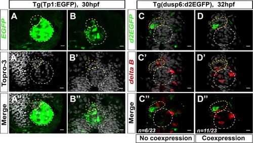
Components of the Notch pathway are mosaically expressed in the parapineal.(A–B) Confocal sections showing the expression of Tg(Tp1bglob:EGFP) transgene (green, A, B), Topro-3 nuclear staining (gray, A’–B’) and merge (A’’–B’’) in the epithalamia of two embryos at 30 hpf (n = 3/19). Notch reporter transgene is strongly expressed in the epiphysis (white circle) and weakly detected in the parapineal (yellow circle) of few embryos (n = 3/19). Embryo view is dorsal, anterior is up; scale bar = 10 µm. Data corresponds to three experiments. (C–D’’) Confocal sections showing the expression of Tg(dusp6:d2EGFP) transgene (green, C, D) and deltaB (red, C’–D’) and merge (C’’–D’’) in the epithalamia of two representative embryos at 32 hpf. deltaB mRNA is detected in only one or two parapineal cells in most embryos (n = 17/23); staining is either co-expressed with Tg(dusp6:d2EGFP) (n = 11/23) or detected in Tg(dusp6:d2EGFP) negative cells (n = 6/23); in n = 6/23, deltaB was not detected in the parapineal. Embryo view is dorsal, anterior is up; epiphysis (white circle), parapineal (yellow circle); scale bar = 10 µm. Data are representative of two experiments.
|