
Figure 1 Supplement 3
- ID
- ZDB-IMAGE-191230-1646
- Publication
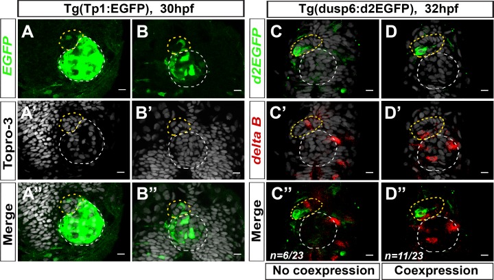
- Wei et al., 2019 - Notch signaling restricts FGF pathway activation in parapineal cells to promote their collective migration
- All Figures
- Figures for Wei et al., 2019
|
Figure 1 Supplement 3
(