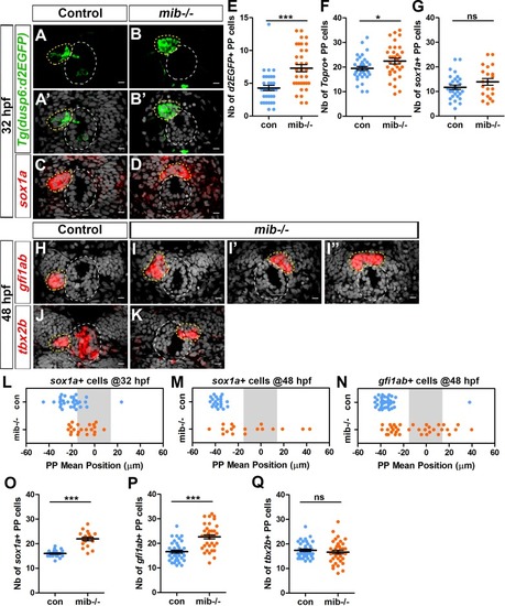
|
<italic>rbpja/b</italic> morphants phenocopy <italic>mindbomb</italic> mutants.(A–B’) Confocal sections showing the expression of gfi1ab (red) at 48 hpf in control embryos (A, n = 33) and in two representative embryos injected with 4 ng of rbpja/b morpholinos (MO) (B, B’); sections are merged with nuclear staining (gray) to highlight the epiphysis (white circle) and parapineal (yellow circle). Embryo view is dorsal, anterior is up; scale bar = 10 µm. (C–D) Dot plots showing the number of Tg(dusp6:d2EGFP) positive parapineal cells (C) and the mean position of parapineal cells highlighted by nuclei staining (Topro-3) (D) at 32 hpf in embryos injected with 4 ng rbpja/b MO (orange dots; n = 36) or in controls (blue dots; n = 28). (E–F) Dot plots showing the mean position (E) or the number (F) of gfi1ab expressing parapineal cells in non-injected controls (blue dots, n = 33) or in rbpja/b MO injected embryos (orange dots, n = 46) at 48 hpf. Both the migration (distance from the midline; p-value=0.0001 in Welch t-test on absolute value) and laterality (left orientation of migration; p-value=0.0002 on Wilcoxon test) are affected in rbpja/b morphants (E). The number of gfi1ab+ cells is also increased in rbpja/b morphants (F, *** p-value<0.0001; Welch t-test). Data are representative of three (A–B’, E–F) or two experiments (C–D).
|