
Figure 5—figure supplement 1.
- ID
- ZDB-FIG-190723-2686
- Publication
- Jiang et al., 2019 - A conserved morphogenetic mechanism for epidermal ensheathment of nociceptive sensory neurites
- Other Figures
-
- Fig. 1
- Fig. 1-S1
- Fig. 1-S2
- fig2
- fig3
- Figure 2—figure supplement 1.
- Figure 3—figure supplement 1.
- Figure 4
- Figure 4—figure supplement 1.
- Figure 4—figure supplement 2.
- Figure 4—figure supplement 3.
- Figure 5
- Figure 5—figure supplement 1.
- Figure 5—figure supplement 2.
- Figure 5—figure supplement 3.
- Figure 6
- Figure 6—figure supplement 1.
- Figure 6—figure supplement 3.
- Figure 7—figure supplement 1.
- All Figure Page
- Back to All Figure Page
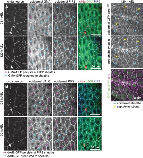
Time-lapse images show dual labeling of epidermal sheaths by PLCδ-PH-Cerulean and GMA-GFP ( |