
Fig. S6
- ID
- ZDB-FIG-181003-25
- Publication
- Yakulov et al., 2018 - CXCL12 and MYC control energy metabolism to support adaptive responses after kidney injury
- Other Figures
- All Figure Page
- Back to All Figure Page
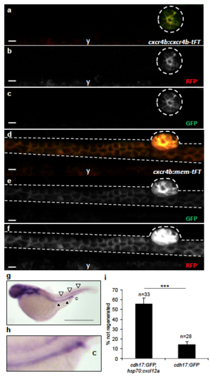
Low-level expression of cxcr4b in pronephric duct cells during normal collective cell migration. (a-c) Using the cxcr4b:cxcr4b-TFT (GFP = short life-time, RFP = long life-time) zebrafish line, lifetime Cxcr4b was only detectable in the corpuscle of Stannius (outlined) at 48 hpf, but not in the pronephros. (d-f) The membrane-resident version cxcr4b:mem-TFT, which does not respond to Cxcl12a signaling and therefore accumulates at the plasma membrane, was detectable in the pronephros (y, yolk) (Scale bars, 10 μm). (g) In situ hybridization depicts expression of cxcl12a in the posterior lateral line (white arrowheads) and in the pronephros (black arrowheads) in zebrafish embryos at 2 dpf (Scale bar, 800 μm). A magnification of the posterior pronephric duct is shown in (h) (c, cloaca). (i) Ectopic expression of Cxcl12a, triggered by heat-shock in the double transgenic cdh17:GFP; hsp-70:cxcl12a zebrafish line, significantly reduces the ability to repair a pronephros injury (means ± SEM; ***, p < 0.001; t-test) . |
| Fish: | |
|---|---|
| Condition: | |
| Observed In: | |
| Stage: | Long-pec |