
Fig. 2
- ID
- ZDB-FIG-180827-14
- Publication
- Ferrero et al., 2018 - Embryonic Microglia Derive from Primitive Macrophages and Are Replaced by cmyb-Dependent Definitive Microglia in Zebrafish
- Other Figures
- All Figure Page
- Back to All Figure Page
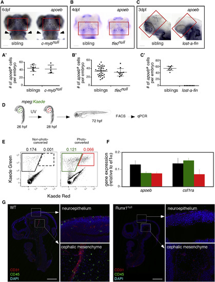
pMFs Are the Unique Source of MG in the Vertebrate Embryo (A–C) Expression of the MG marker apoeb (red quadrants) in the brains of cmybnull (A), tfecnull (B), alk8null(lost-a-fin) (C), and their sibling control animals at the indicated developmental stages. For (A), cmybnull embryos are identified by the concomitant absence of lymphoid-specific rag1 (arrowheads) transcripts in the thymus. Scale bar: 50 μm. (A′–C′) Quantification of apoeb+ MG in embryos of indicated genotypes. Each symbol represents a single embryo. Error bars represent mean ± SEM. (D) Strategy for transient fate mapping of pMFs in vivo. (E) Flow cytometry showing absence of kaede-Red expression in non-photoconverted embryos (left panel), whereas cells expressing both forms of kaede were present and sortable in the photoconverted embryos (right panel). (F) qPCR analyses for apoeb and csf1ra on mpeg1:kaede-Green cells sorted from control and photoconverted embryos, and on mpeg1:kaede-Green/Red cells from photoconverted embryos. The color code matches the gates on the FACS plots. Error bars represent mean ± SD (n = 3 experiments). (G) Sagittal sections of E10.5 Runx1null embryos and their littermates (upper body) immunostained for CD31 (vascular) and CD45 (hematopoietic) markers, with DAPI used as a nuclear counterstain. Tile-scanned images show that Runx1null mutants are completely devoid of CD45+ MG in the neuroepithelium and cephalic mesenchyme (n = 3). Scale bar: 500 μm. See also Figures S1–S3. |
| Genes: | |
|---|---|
| Fish: | |
| Anatomical Terms: | |
| Stage Range: | Protruding-mouth to Day 6 |
| Fish: | |
|---|---|
| Observed In: | |
| Stage Range: | Protruding-mouth to Day 6 |