
Fig. 2
- ID
- ZDB-IMAGE-180827-12
- Genes
- Publication
- Ferrero et al., 2018 - Embryonic Microglia Derive from Primitive Macrophages and Are Replaced by cmyb-Dependent Definitive Microglia in Zebrafish
- All Figures
- Figures for Ferrero et al., 2018
|
Fig. 2
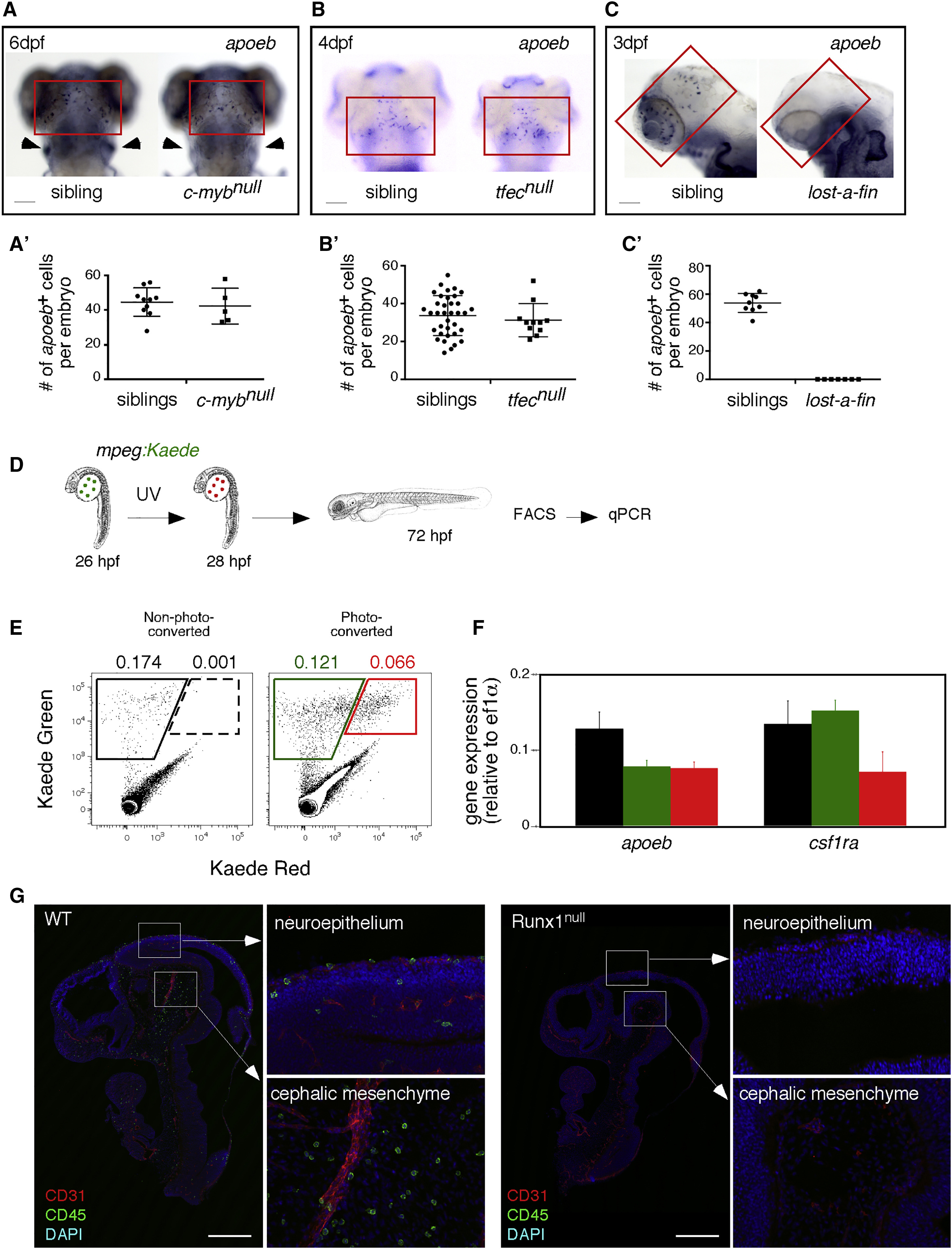
pMFs Are the Unique Source of MG in the Vertebrate Embryo
(A–C) Expression of the MG marker apoeb (red quadrants) in the brains of cmybnull (A), tfecnull (B), alk8null(lost-a-fin) (C), and their sibling control animals at the indicated developmental stages. For (A), cmybnull embryos are identified by the concomitant absence of lymphoid-specific rag1 (arrowheads) transcripts in the thymus. Scale bar: 50 μm.
(A′–C′) Quantification of apoeb+ MG in embryos of indicated genotypes. Each symbol represents a single embryo. Error bars represent mean ± SEM.
(D) Strategy for transient fate mapping of pMFs in vivo.
(E) Flow cytometry showing absence of kaede-Red expression in non-photoconverted embryos (left panel), whereas cells expressing both forms of kaede were present and sortable in the photoconverted embryos (right panel).
(F) qPCR analyses for apoeb and csf1ra on mpeg1:kaede-Green cells sorted from control and photoconverted embryos, and on mpeg1:kaede-Green/Red cells from photoconverted embryos. The color code matches the gates on the FACS plots. Error bars represent mean ± SD (n = 3 experiments).
(G) Sagittal sections of E10.5 Runx1null embryos and their littermates (upper body) immunostained for CD31 (vascular) and CD45 (hematopoietic) markers, with DAPI used as a nuclear counterstain. Tile-scanned images show that Runx1null mutants are completely devoid of CD45+ MG in the neuroepithelium and cephalic mesenchyme (n = 3). Scale bar: 500 μm.
See also Figures S1–S3.