
Fig. S2
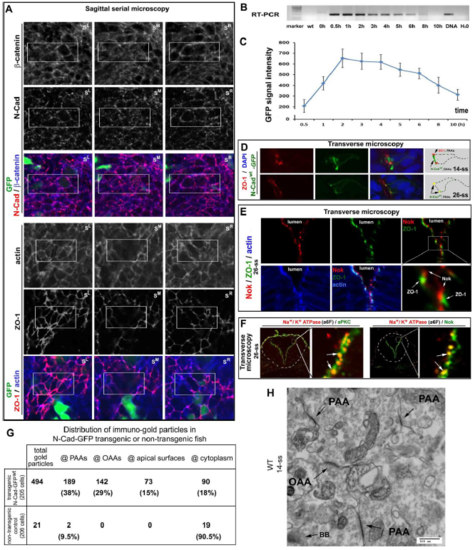
The development of the PAAs and OAAs and their dynamic molecular compositions A. GFP-assisted sagittal serial microscopy revealed that N-Cad and -catenin but not ZO-1 or actin localized to the opposing apical surfaces. The boxed areas are presented in Fig. 2A at higher magnifications. B. An RT-PCR revealed that the mRNA products of the N-Cadwt-GFP transgene reached its highest level 30 min after a heat shock and disappeared completely by 10 hours after the heat shock. C. The N-Cadwt-GFP fluorescence intensities of whole embryos reached the highest level 2 hours after the heat shock, and fell by 77% 10 hours after the heat shock. The fluorescence intensities were measured with Q-imaging under a stereo-fluorescence scope (ten embryos; means ± SEM). D. Single channel and merged images of Fig. 2B are presented to better illustrate the distributions of N-Cadwt-GFP to the PAAs and OAAs; N-Cadwt GFP was transiently-induced by heat shock from a transgenic construct injected into the embryos. Note that N-Cadwt-GFP enriched at both the PAAs and OAAs at 14-ss, but only at the PAAs at 26-ss. Weaker N-Cadwt-GFP signals persisted on the lateral membranes. The drawings summarize the distributional dynamics of ZO-1 and N-Cad-GFP. E. Nok, a cytoplasmic partner of Crb proteins, closely associated with the apical side of ZO-1 sites. F. Na+/K+ ATPase colocalized with aPKC and Nok at 26 ss (arrows), although Nok and aPKC distributed more broadly than Na+/K+ ATPase did. G. The table presents the subcellular distributions of immuno-EM gold particles in the early neural rods (at 14-ss) of N-Cad-GFPwt-expressing transgenic embryos and non-transgenic wildtype control embryos: The numbers of gold particles (as well as their corresponding percentages in total particles) distributed at the PAAs, OAAs, opposing apical surface, and cytoplasm are tabulated. Note the enrichment of gold particles at the OAAs and PAAs in N-Cad-GFPwt-expressing embryos but not in wildtype control embryos. H. An immuno-EM micrograph of the midline region of a wildtype neural rod revealed that the PAAs and OAAs survived the Triton X-100 extraction of the staining procedure and that no gold particles were observed at the PAAs and OAAs. |