
FIGURE
Fig. 7
- ID
- ZDB-FIG-180821-19
- Publication
- McMillan et al., 2018 - A regulatory pathway involving retinoic acid and calcineurin demarcates and maintains joint cells and osteoblasts in the fin regenerate
- Other Figures
- All Figure Page
- Back to All Figure Page
Fig. 7
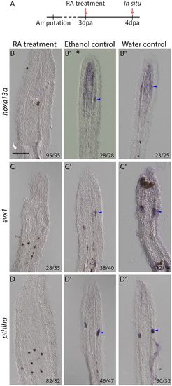
RA treatment leads to the inhibition of joint cell marker expression. (A) Fish were treated with RA from 3 to 4 dpa. (B-D) ISH on longitudinal cryosections at 4 dpa/1 dot indicate hoxa13a (B), evx1 (C) and pthlha (D) expression are lost in joint cells of RA-treated fish. (B′-D″) Expression remains in joint cells (blue arrowheads) of ethanol (B′-D′) and water (B″-D″) controls. Numbers in each panel represent the number of sections with the expression pattern over the total number of sections analyzed. Scale bar: 100 μm (in B for B-D″). |
Expression Data
| Genes: | |
|---|---|
| Fish: | |
| Conditions: | |
| Anatomical Term: | |
| Stage: | Adult |
Expression Detail
Antibody Labeling
Phenotype Data
Phenotype Detail
Acknowledgments
This image is the copyrighted work of the attributed author or publisher, and
ZFIN has permission only to display this image to its users.
Additional permissions should be obtained from the applicable author or publisher of the image.
Full text @ Development