
Fig. 1
- ID
- ZDB-FIG-180821-13
- Publication
- McMillan et al., 2018 - A regulatory pathway involving retinoic acid and calcineurin demarcates and maintains joint cells and osteoblasts in the fin regenerate
- Other Figures
- All Figure Page
- Back to All Figure Page
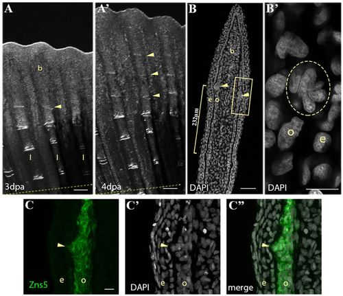
Periodic formation of a cluster of joint-forming cells during fin regeneration. (A-A′) Bright-field images of fin regenerate at 3 dpa (A) and 4 dpa (A′) illustrate the periodic addition of joints (yellow arrowheads) to the distal end of the rays. Dashed yellow line indicates amputation plane. (B) DAPI staining on a 4 dpa regenerate longitudinal cryosection illustrates a cluster of nuclei (yellow arrowheads) 232 μm (yellow bracket) from a mature joint. (B′) Magnification of the boxed area in B showing the nuclei cluster (yellow circle). (C) Zns5 immunohistostaining labels the joint cell cluster and adjacent osteoblasts. (C′) DAPI staining of the same section showing the cell cluster nuclei. (C″) Merged image of C and C′. b, blastema; e, epidermis; l, lepidotrichia; o, osteoblast. Scale bars: 200 μm (A,A′); 50 μm (B); 10 μm (B′; in C for C-C″). |
| Antibody: | |
|---|---|
| Fish: | |
| Condition: | |
| Anatomical Terms: | |
| Stage: | Adult |