
Fig. 4
- ID
- ZDB-FIG-180821-16
- Publication
- McMillan et al., 2018 - A regulatory pathway involving retinoic acid and calcineurin demarcates and maintains joint cells and osteoblasts in the fin regenerate
- Other Figures
- All Figure Page
- Back to All Figure Page
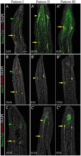
Sequential activation of hoxa13a, evx1 and pthlha expression in the presumptive joint cells. Double FISH and DAPI counterstains on longitudinal cryosections of 4 dpa fin regenerates illustrate three distinct patterns of expression. (A-C′) Patterns I and II are observed in rays with presumptive joint cells (arrowheads). (A″-C″) Pattern III is observed in rays without presumptive joint cells. (A-C″) In joint-forming cells (yellow arrows), the three markers are co-expressed in the joint-forming cells. (A,A′,C,C′) In the presumptive joint cells (green or yellow arrowheads), hoxa13a is expressed alone (Pattern I, A,C) or co-expressed with evx1 (Pattern II, A′) or pthlha (Pattern II, C′). (B′,C′) pthlha is also co-expressed with evx1 (B′). (B,B′) evx1 is either expressed alone (B) or co-expressed with pthlha (B′). Numbers in each panel represent the number of sections with the expression pattern over the total number of sections analyzed. Scale bar: 50 μm (in A for A-C″). |
| Genes: | |
|---|---|
| Fish: | |
| Condition: | |
| Anatomical Term: | |
| Stage: | Adult |