Fig. 6-S1
- ID
- ZDB-FIG-180417-30
- Publication
- Rogers et al., 2017 - Nodal patterning without Lefty inhibitory feedback is functional but fragile
- Other Figures
- All Figure Page
- Back to All Figure Page
|
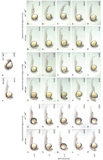
Artificial, uniform Nodal inhibition rescues morphology at 1 dpf in lefty1-/-;lefty2-/- mutants. Wild type and lft1-/-;lft2-/- mutant embryos were exposed to 0, 1.25, 2.5, 3.75, 5, or 6.25 μM SB-505124 (Nodal inhibitor drug) starting at the 8 cell stage, sphere stage, or shield stage. Drug was removed at 24 hr post-fertilization. Rescue at earlier stages requires lower concentrations (2.5–3.75 μM) of SB-505124 than rescue at later stages (5–6.25 μM). (A–B) Unexposed wild type (A) and lft1-/-;lft2-/- mutant (B) embryos at 1 dpf. (C–Q’) Embryos exposed to SB-505124 from the 8 cell stage to 24 hpf (C–G’), sphere stage to 24 hpf (H–L’), and shield stage to 24 hpf (M–Q’) at 1 dpf. Number of embryos with same phenotype as image/total is indicated in bottom right of images. Cyclopia and loss of head and trunk mesendoderm are typical Nodal loss-of-function phenotypes (Gritsman et al., 1999; Hagos and Dougan, 2007), and can be observed in both wild type and mutant embryos treated with higher amounts of SB-505124 (e.g., F and F’). Wild type embryos exposed to 3.75 μM at the 8 cell stage (E) exhibited cyclopia (15/27, shown here) and normal morphology (12/27). Wild type embryos exposed to 3.75 μM at sphere stage (J) exhibited mild cyclopia (6/30), moderate cyclopia (13/30, shown here), necrosis and cyclopia (1/30), normal morphology (9/30), and 1/30 died. Some images shown here are also used in Figure 6. |
| Fish: | |
|---|---|
| Condition: | |
| Observed In: | |
| Stage: | Prim-5 |

