FIGURE

Fig. 1-s1

ID
ZDB-FIG-180403-53
Publication
Norris et al., 2017 - Toddler signaling regulates mesodermal cell migration downstream of Nodal signaling
Other Figures
All Figure Page
Back to All Figure Page
Fig. 1-s1

Initial specification of endoderm is normal in toddler mutants.

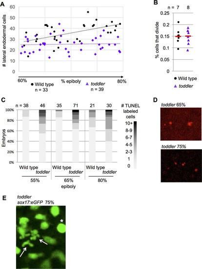
(A–C) n = number of embryos (A) Number of lateral endodermal cells in wild-type and toddler mutant embryos during gastrulation. (B) Rate of endodermal cell division in wild-type and toddler mutant embryos. 40–60 cells were tracked per embryo and the percentage of those cells that divided is shown. Red bars are averages. (C) Percentage of embryos with a given number of TUNEL +cells on one side of the embryo. (D) Representative images of TUNEL staining at two time points in toddler mutants. (E) Live tracking of endoderm using sox17:GFP transgenic fish captures two endodermal cells undergoing cell death during gastrulation in toddler mutants (arrows). A cell that is about to divide is also present (asterisk).

Expression Data
Gene:
Fish:
Anatomical Term:
Stage: 75%-epiboly

Expression Detail
Antibody Labeling
Phenotype Data
Fish:
Observed In:
Stage Range: Shield to 75%-epiboly

Phenotype Detail
Acknowledgments
This image is the copyrighted work of the attributed author or publisher, and ZFIN has permission only to display this image to its users. Additional permissions should be obtained from the applicable author or publisher of the image. Full text @ Elife