
Fig. 6
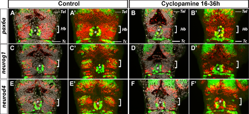
Hh signalling is required for the expression of pax6a, neurog1 and neurod4 in the Habenular nuclei. Confocal sections (A-F) or 15μm maximum projections (A’-F’) showing the heads of control treated embryos (A-A’, n = 9; C-C’, n = 6; E-E’, n = 6) or those treated from 16 hpf with cyclopamine (B-B’, n = 10, D-D’, n = 7, F-F’, n = 6) after a whole-mount in situ hybridization against pax6a (A,A’,B,B’), neurog1 (C,C’,D,D’) and neurod4 (E,E’,F,F’) (red) and immunostaining against HuC/D protein (green); cell nuclei staining (grey) makes visible brain structures in the confocal sections (A-F). The overall morphology of the head appears normal in cyclopamine treated embryos and the expression of HuC/D does not appear to be affected in the telencephalon (Tel), in the epiphysis (*) nor in the tectum (Tc). In contrast, HuC/D expression is absent or strongly reduced in the habenular domain (Hb, white brackets) of cyclopamine treated embryos. The expression of pax6a is strongly reduced (B-B’, n = 10/10) in the habenular domain (Hb, white brackets) of cyclopamine treated embryos. The expression of neurog1 and neurod4 is also abrogated specifically in the habenular domain of cyclopamine treated embryos (D-D’, n = 7/7, F-F’, n = 6/6). All embryos are at 36 hpf. Embryos are viewed dorsally with anterior up. |
| Genes: | |
|---|---|
| Antibody: | |
| Fish: | |
| Condition: | |
| Anatomical Terms: | |
| Stage: | Prim-25 |
| Fish: | |
|---|---|
| Condition: | |
| Observed In: | |
| Stage: | Prim-25 |