
Fig. 5
- ID
- ZDB-FIG-170706-20
- Publication
- Pei et al., 2016 - Additive reductions in zebrafish PRPS1 activity result in a spectrum of deficiencies modeling several human PRPS1-associated diseases
- Other Figures
- All Figure Page
- Back to All Figure Page
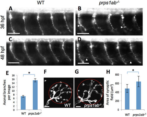
prps1a:prps1b double mutants display abnormal morphology in primary motor neuron and neuromast hair cell innervation. (A–D) Primary motor neurons stained using the znp-1 antibody. (A,B) Morphology of motor neuron axons in wild-type (A) and double mutant (B) embryos at 36 hpf. White arrowheads point to aberrant pseudopodia. The axons in the double mutants were shorter with increased branching compared to wild-type siblings. (C,D) Morphology of motor neuron axons in wild-type (C) and double mutant (D) embryos at 48 hpf. White arrowheads point to elongated branches. 10 embryos are used for each group of staining, with one representative image shown. (E) Quantification of axon branching. Y-axis indicates the number of elongated branches per image that frames 5 neurons. The increase in axon branching is significant in the double mutant (n = 10, p < 0.001). (F–H) Afferent neurons innervating neuromast hair cells stained with a calretinin antibody. (F,G) Morphology of innervating afferent neurons and fibers in wild-type (F) and double mutant (G) at 6 dpf, red circles are examples of the area calculated. (H) Quantification of the innervating areas. The analysis was done with 12 neuromasts from 9 wild-type embryos and 13 neuromasts from 6 double mutants. The area each neuron innervates is significantly greater in the double mutants. The difference between wild-type and double mutant is significant (Tukey's multiple comparisons, p < 0.01). Scale bars: 50 μm in (A–D); 5 μm in (F,G). |
| Fish: | |
|---|---|
| Observed In: | |
| Stage Range: | Prim-25 to Day 6 |