
Fig. 4
- ID
- ZDB-FIG-170706-19
- Publication
- Pei et al., 2016 - Additive reductions in zebrafish PRPS1 activity result in a spectrum of deficiencies modeling several human PRPS1-associated diseases
- Other Figures
- All Figure Page
- Back to All Figure Page
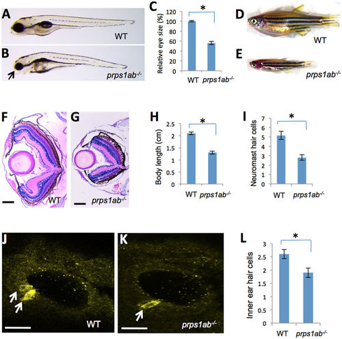
prps1a;prps1b double mutant displays defects in the eye, craniofacial structure, general growth, neuromast and inner ear hair cells. (A,B) Eye phenotype of the double mutant. Images show genetically-related wild-type (A) and the double mutant (B) at 5 dpf. Arrow in B points to the small eye and abnormal craniofacial structure of the double mutant. Most double mutants fail to inflate the swim bladder. (C) Quantification of the reduction in the eye size. Eye area is calculated using Image J. The reduction is significant (n = 9, p < 0.001). (D,E) Morphology of wild-type (D) and the double mutant fish (E) at 35 dpf, only 5% of the double mutant fish survive to this age. (F,G) Retinal lamination of wild-type (F) and the double mutant (G) at 35 dpf. The double mutant had a poorly formed retinal inner plexiform layer and has an under-developed retinal pigment epithelial layer. (H) Quantification of the body length at 35 dpf. The reduction in the double mutant is significant (n = 6, p < 0.001). (I) Quantification of the neuromast hair cells in the double mutants at 3 dpf. The reduction in the double mutant is significant (n = 9 for wild-type, n = 11 for the double mutant, p < 0.001). (J,K) Inner ear hair cells in the embryos at 32 hpf were stained with a mixture of myosin-VIIa and hair cell soma-1 antibodies. 14 embryos per group were used for inner ear hair cell analysis. Representative images are shown. White arrows indicate positive staining of hair cells. (L) Quantification of the reduction of inner ear hair cells. The reduction in the double mutants is significant (n = 14, p = 0.003). For all graphs, the average and s.e.m are shown. prps1ab−/− indicates the prps1a:prps1b double mutants. Scale bars: 200 μm in (F,G); 50 μm in (J,K). |
| Fish: | |
|---|---|
| Observed In: | |
| Stage Range: | Prim-15 to Days 30-44 |