
Fig. 1 S2
- ID
- ZDB-FIG-170310-3
- Publication
- Bloomekatz et al., 2017 - Platelet-derived growth factor (PDGF) signaling directs cardiomyocyte movement toward the midline during heart tube assembly
- Other Figures
- All Figure Page
- Back to All Figure Page
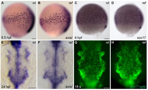
The anterior endoderm forms normally in ref mutant embryos. (A–D) Lateral views, dorsal to the right, depict the expression of axial at 8.5 hpf (A,B) or sox17 at 8 hpf (C,D). The number and distribution of endoderm precursor cells is similar in wt (A,C) and ref mutant (B, n = 8/8; D, n = 14/14) embryos during gastrulation stages. (E–H) Dorsal views, anterior to the top, depict the anterior endoderm, visualized with the expression of axial at 24 hpf (E,F) or the endodermal reporter transgene Tg(sox17:egfp) at 18 s (G,H). The width and morphology of the anterior endoderm is similar in wt (E,G) and ref mutant (F, n = 13/13; H, n = 7/7) embryos during the stages when cardiac fusion takes place. Scale bars: 60 μm. |