
Fig. 1 S1
- ID
- ZDB-FIG-170310-2
- Publication
- Bloomekatz et al., 2017 - Platelet-derived growth factor (PDGF) signaling directs cardiomyocyte movement toward the midline during heart tube assembly
- Other Figures
- All Figure Page
- Back to All Figure Page
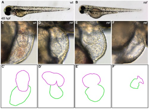
ref mutant embryos display a range of cardiac defects. (A,B) Lateral views of live wild-type (wt, A) and ref mutant (B) embryos at 48 hpf. ref mutants exhibit a mild pericardial edema. (C–F) Lateral views from the right side of wt (C) and ref mutant (D–F) hearts reveal a variety of abnormal cardiac morphologies in ref mutants. The most commonly observed phenotype was a bifurcated ventricle (D), some other embryos exhibited abnormal looping (E) or a severely shrunken heart (F), and a few embryos had cardia bifida (not shown). See Tables 1 and 2 for more information regarding the incomplete penetrance and variable expressivity of the ref mutant phenotype. (C'–F') Cartoons outline the cardiac morphologies shown in C–F, with the ventricle in magenta and the atrium in green. |