
Fig. 1
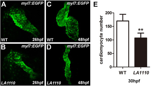
LA1110 mutants exhibit a reduction in cardiomyocyte cell number. (A and B) Tbx20 mutant embryos (B) form a thin and small heart tube at 26hpf compared with that of wild-type siblings (A), as indicated by the fluorescent labeling of differentiated cardiomyocytes by the myl7:EGFP transgene. (C and D) Wild type embryos display a looped two-chambered heart at 48hpf (C), whereas LA1110 mutant embryos (D) exhibit a thin, stretched and string-like heart. (E) Graph of the average number of GFP-positive cardiomyocytes in wild type and LA1110 mutant embryos at 30hpf (n=5). Error bars indicate standard deviations. Asterisks indicate a significant difference (**p<0.01). |
| Gene: | |
|---|---|
| Fish: | |
| Anatomical Term: | |
| Stage Range: | Prim-5 to Long-pec |
| Fish: | |
|---|---|
| Observed In: | |
| Stage Range: | Prim-5 to Long-pec |
Reprinted from Developmental Biology, 421(2), Lu, F., Langenbacher, A., Chen, J.N., Tbx20 drives cardiac progenitor formation and cardiomyocyte proliferation in zebrafish, 139-148, Copyright (2017) with permission from Elsevier. Full text @ Dev. Biol.