
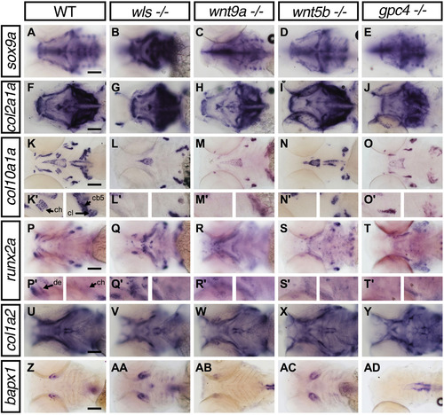
Fig. 7
Lower jaw gene expression pattern of chondrocyte and bone markers inwls,wnt9a,wnt5bandgpc4mutants. Whole-mount RNA in situ hybridization of chondrocyte differentiation markers; sox9a (A-E) and chondrocyte matrix; col2a1 (F-J). Hypertrophic chondrocyte marker; col10a1a (K-O), Bone and tendon marker; col1a2 (P-T), osteoblastic progenitor marker; runx2a (U-Y) and joint marker; bapx1 (Z-AD). (K′-L′) 40X image focused on ceratohyal (ch; left) and both cleithrum and ceratobranchial 5 (cl, cb; right). (P′-T′) 40X images focused on dentary (de; left) and ceratohyal (right). Apparent increase in sox9a, runx2a expression pattern in wls -/- with concomitant loss of col10a1a. Black arrowhead points to the position of Meckel's cartilage. Scale=50 µm.. |
| Genes: | |
|---|---|
| Fish: | |
| Anatomical Terms: | |
| Stage: | Day 4 |
| Fish: | |
|---|---|
| Observed In: | |
| Stage: | Day 4 |
Reprinted from Developmental Biology, 421(2), Ling, I.T., Rochard, L., Liao, E.C., Distinct requirements of wls, wnt9a, wnt5b and gpc4 in regulating chondrocyte maturation and timing of endochondral ossification, 219-232, Copyright (2017) with permission from Elsevier. Full text @ Dev. Biol.