
Fig. 6
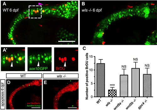
Chondrocyte proliferation defect inwlsmutant precedes the differentiation defect. (A, B) Representative BrdU assay confocal stacked image in WT 6 dpf (A) and wls -/- (B) embryo with zoomed views of WT BrdU (red) colocalizing with chondrocytes (A’) in the Meckel's cartilage (green). Arrowhead points to a representative chondrocyte that expressed both sox10 and BrdU and used for quantification. (C) Quantification of BrdU positive cells in Wnt mutants. Proliferation significantly differed in wls -/- compared to WT (****p<0.001; Kruskal-Wallis test and Dunn's multiple comparison test, NS = not significant). (D, E) No difference in chondrocyte apoptosis as showed with Acridine orange live staining at 6 dpf in wls -/-. Scale=50 µm. |
| Fish: | |
|---|---|
| Observed In: | |
| Stage: | Day 6 |
Reprinted from Developmental Biology, 421(2), Ling, I.T., Rochard, L., Liao, E.C., Distinct requirements of wls, wnt9a, wnt5b and gpc4 in regulating chondrocyte maturation and timing of endochondral ossification, 219-232, Copyright (2017) with permission from Elsevier. Full text @ Dev. Biol.