
FIGURE
Fig. S2
- ID
- ZDB-FIG-170203-10
- Publication
- Ling et al., 2017 - Distinct requirements of wls, wnt9a, wnt5b and gpc4 in regulating chondrocyte maturation and timing of endochondral ossification
- Other Figures
- All Figure Page
- Back to All Figure Page
Fig. S2
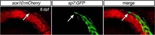
Supplementary material: Supplementary Fig 2. Osterix-expressing cells in 8 dpf WT embryos. 40X Confocal maximum intensity projection images of 8 dpf Meckel's cartilage. Osterix expressing cells are observed at the position of the dentary adjacent to the body of Meckel's above the perichondrium. A single osterix-expression cell (arrow) is observed at the level of the perichondrium indicating the differentiation of perichondral cells to osteoblast to form the future mentomeckelian perichondral bone. |
Expression Data
| Gene: | |
|---|---|
| Fish: | |
| Anatomical Term: | |
| Stage: | Days 7-13 |
Expression Detail
Antibody Labeling
Phenotype Data
Phenotype Detail
Acknowledgments
This image is the copyrighted work of the attributed author or publisher, and
ZFIN has permission only to display this image to its users.
Additional permissions should be obtained from the applicable author or publisher of the image.
Reprinted from Developmental Biology, 421(2), Ling, I.T., Rochard, L., Liao, E.C., Distinct requirements of wls, wnt9a, wnt5b and gpc4 in regulating chondrocyte maturation and timing of endochondral ossification, 219-232, Copyright (2017) with permission from Elsevier. Full text @ Dev. Biol.