
Fig. S2
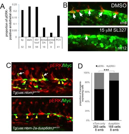
Chemical and genetic blockade of pERK in zebrafish embryos (A) Quantification of pERK-positive endothelial cells (i.e. also positive for fli1:egfp) in trunk vessels of wild type Tg(fli1a:egfp)y1 zebrafish embryos at 24 hpf. Graph shows average proportion of the total number of pERK-positive endothelial cells that contribute to indicated blood vessel. Numbers indicate total numbers of pERK-positive endothelial cells counted in 16 embryos. Hematopoietic cells, as identified by their round morphology, were not counted in this assessment. (B) pERK immunostaining of Tg(fli1a:egfp)y1 embryos following treatment for 30 minutes at 20 hpf with 0.1% DMSO or 15 μM SL327. Lateral views, anterior is left, dorsal is up. pERK-positive endothelial cells in the dorsal aorta are indicated by arrows. (C) Embryos bearing indicated transgenes at 21 hpf immunostained for pERK (red) and the nuclear localized Myc epitope (green) on tdTomato (nTom). White arrows denote co-staining of Myc and pERK, yellow arrows indicate Myc-positive;pERK-negative cells. (D) Quantification of pERK immunostaining in cells expressing indicated transgene. Number of cells and embryos used for analysis is indicated. ***p<0.001. |