Fig. S6
- ID
- ZDB-FIG-160927-42
- Publication
- Liu et al., 2016 - Fscn1 is required for the trafficking of TGF-β family type I receptors during endoderm formation
- Other Figures
- All Figure Page
- Back to All Figure Page
|
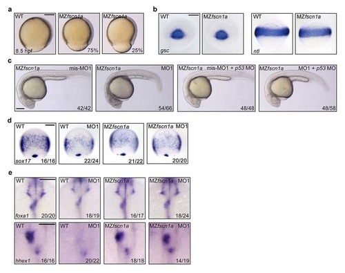
MZfscn1a mutants exhibit defects in epiboly progression and endoderm development, but have normal mesoderm formation. (a) Representative bright-field images of wild-type and MZfscn1a mutants at 8.5 hpf. The percentage of MZfscn1a mutants with various degrees of epiboly defects was shown. Scale bar, 200 µm. (b) Wild-type and MZfscn1a mutant embryos were harvested at the shield stage for in situ hybridization with gsc and ntl probes. Scale bar, 200 µm. (c) Representative bright-field images of MZfscn1a mutant embryos injected with indicated 4 ng fscn1a MOs together with or without 4 ng p53 MO at 24 hpf. Scale bar, 200 µm. (d) The expression of sox17 at 75% epiboly stage was examined in wild-type and MZfscn1a mutant embryos injected with indicated fscn1a MOs. Note that no visible enhancement of endodermal related defects was observed in MO1 injected MZfscn1a embryos compared with uninjected mutants. Scale bar, 200 µm. (e) The expression of foxa1 and hhex1 at 28 hpf was examined in wild-type and MZfscn1a mutant embryos injected with indicated fscn1a MOs. Note that the liver and pancreatic buds were normally formed in MO1 injected MZfscn1a embryos compared with uninjected mutants. Scale bar, 200 µm in the upper panels and 100 µm in the lower panels. |
| Genes: | |
|---|---|
| Fish: | |
| Knockdown Reagent: | |
| Anatomical Terms: | |
| Stage Range: | Shield to Prim-5 |
| Fish: | |
|---|---|
| Knockdown Reagent: | |
| Observed In: | |
| Stage Range: | 75%-epiboly to Prim-5 |

