
FIGURE
Fig. 7
- ID
- ZDB-FIG-160606-9
- Publication
- Sepich et al., 2016 - Intracellular Golgi Complex Organization Reveals Tissue Specific Polarity during Zebrafish Embryogenesis
- Other Figures
- All Figure Page
- Back to All Figure Page
Fig. 7
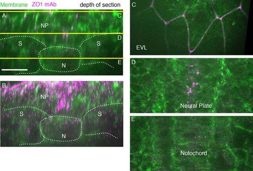
ZO1 antibody-labeled tight junctions in 5 somite stage wild-type embryo (11.7 hpf). A: A single-transverse-plane ZO1 antibody (magenta) cell membranes (green, Transgene [actb::Myosinl12-GFP]). Yellow lines show depth of sections in C,D,E. Dorsal is to the top. Lines mark tissue boundaries. B: Transverse z-projected section of wild-type embryo (spt sibling). C: EVL cells. D: Ventral neural plate. E: Notochord. N, notochord; NP, neural plate; S, somites. Scale bars = 20 µm. |
Expression Data
Expression Detail
Antibody Labeling
Phenotype Data
Phenotype Detail
Acknowledgments
This image is the copyrighted work of the attributed author or publisher, and
ZFIN has permission only to display this image to its users.
Additional permissions should be obtained from the applicable author or publisher of the image.
Full text @ Dev. Dyn.