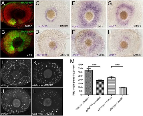
Pharmacological activation of the RA pathway in wild-type embryos mimics the gdf6a mutant eye phenotype. (A,B). Representative lateral views of maximum intensity projections of eyes from Tg[RARE:YFP] embryos immunostained for GFP (RARE:YFP, green) and counterstained with DAPI (red). Eyes are from embryos that were incubated in vehicle control (DMSO; A) or in 13-cis RA (B), exposed to a pulse of UV light, and then fixed and immunostained at 33hpf. The majority of RPCs in B have responded to RA by activating the RARE:YFP transgene. (C-H) Lateral views of eyes from wild-type embryos treated with vehicle (DMSO; C,E,G) or the RAR agonist AM580 (25nM; D,F,H) from 24 to 36hpf and analysed at 60hpf for expression of CMZ genes col15a1b (C,D), ccnd1 (E,F) and atoh7 (G,H). (I-L) Representative lateral views of maximum intensity projections of 60hpf embryonic eyes immunostained for PH3. Eyes from an untreated sibling and untreated gdf6au768 mutant (I and J, respectively) and from a DMSO-treated wild type and an AM580-treated wild type (K and L, respectively) are shown. (M) Graph quantifying PH3+ cells in I-L. The average number of PH3+ cells per retina (n=10 embryos) was calculated and then plotted with standard error bars (95% confidence limits; pair-wise comparisons using Student′s t-test, ***P≤0.0001).
|
