
Fig. S1
- ID
- ZDB-FIG-160503-11
- Publication
- Valdivia et al., 2016 - Antagonism between Gdf6a and retinoic acid pathways controls timing of retinal neurogenesis and growth of the eye in zebrafish
- Other Figures
- All Figure Page
- Back to All Figure Page
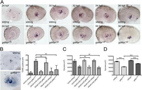
gdf6a mutants exhibit microphthalmia independently of cell death. (A) Lateral views of wild-type (top row) and gdf6au768 mutant (lower row) eyes showing apoptotic cells between 24 hpf - 36 hpf. No obvious difference in numbers is apparent between wild-type and mutants (p≥0.07 for all time points; n≥10 embryos per stage per genotype). (B) Left: lateral views showing apoptotic cells (purple) wildtype (top) and gdf6au900 mutant eyes. Right: Graph showing that the elevated number of TUNEL+ cells (blue) in gdf6au900 mutant eyes at 32 hpf (bottom image) is reduced to near wild-type levels by incubating embryos with 400 µM Z-VAD-FMK caspase inhibitor from 14 hpf - 31 hpf (**Student’s t-test, p<0.05; n ≥ 5 embryos per genotype; NS, no significant difference). (C) Graph showing that gdf6au900 mutants exhibit significantly smaller eyes than their siblings at 3 dpf independently of Z-VAD-FMK treatment (n≥7 embryos; NS, no significant difference). (D) Graph showing that both alleles of gdf6a are associated with smaller eyes, with gdf6au900 mutant eyes smaller than those in gdf6au768 mutants (***Student’s t-test, p<0.0001; n≥5 embryos). |