|
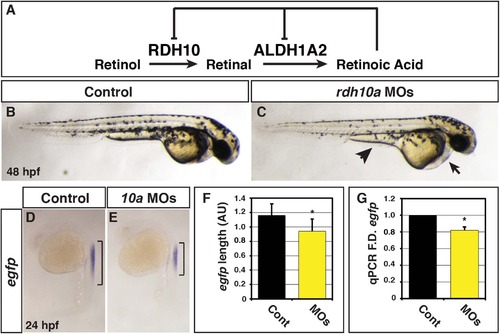
Rdh10a deficient embryos have decreased RA signaling. (A) Schematic of the metabolic pathway that synthesizes RA during development and negative regulation of Rdh10 and Aldh1a2 expression from mice and Xenopus studies [23, 28, 29]. (B) Control sibling embryo. (C) Embryos injected with rdh10a MOs. Arrow indicates pericardial edema. Arrowhead indicates yolk extension. (D-E) ISH for egfp expression in Tg(12XRARE-ef1a:EGFP)sk72 control sibling and Rdh10a deficient embryos. Brackets indicate the length of egfp expression in the spinal cord. (F) Measurements of egfp expression length in the spinal cord using arbitrary units (AU) from in situ hybridization (ISH) of Tg(12XRARE-ef1a:EGFP)sk72 control embryos (n = 16) and Rdh10a deficient embryos (n = 15). (G) RT-qPCR for egfp expression at 24 hpf in control sibling and Rdh10a deficient embryos. In all graphs, asterisks indicate a statistically significant difference (p<0.05) and bars indicate standard deviation.
|