
Fig. S3
- ID
- ZDB-FIG-151022-13
- Publication
- Mateus et al., 2015 - Control of tissue growth by Yap relies on cell density and F-actin in zebrafish fin regeneration
- Other Figures
- All Figure Page
- Back to All Figure Page
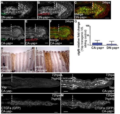
ctgfa is conserved as a direct transcriptional target of Yap in zebrafish. Related to Figs 2 and 3. A-F Representative immunostainings with anti-Yap and anti-RFP in 24 hpa longitudinal sections of CA-yap and DN-Yap positive transgenics, upon heat-shock. A-C DNYap+; D-F CA-Yap+. Note that for further experiments anti-Yap was used instead of anti-RFP due to clearer intracellular labeling. G qPCR determination of ctgfb relative expression levels in CA-yap and DN-yap positive transgenics versus respective sibling controls, upon single heatshock at 72 hpa. Logarithmic scale, base 10. H-I Representative in situ hybridization for ctgfa in 24 hpa fins of CA-yap sibling control (H) and CA-yap+ fish (I). Fins were collected at 2 hpHS. n=3 fins per condition. Scale bars correspond to 500µm. J-M Representative immunostainings with anti-Yap and anti-GFP in 72 hpa longitudinal sections of ctgfa:eGFP; CA-yap double transgenics, fins were fixed at 7 hpHS. J,K CA-yap sibling control; L,M CAyap+. Note that due to high Yap levels in L, normal settings in acquisition of images in J, L had to be decreased to avoid image saturation. n=9 sections, 3 fish per condition. Scale bars correspond to 50µm. Dashed lines indicate amputation plane. |