
Fig. S2
- ID
- ZDB-FIG-151022-12
- Publication
- Mateus et al., 2015 - Control of tissue growth by Yap relies on cell density and F-actin in zebrafish fin regeneration
- Other Figures
- All Figure Page
- Back to All Figure Page
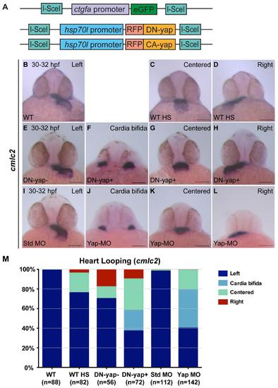
DN-Yap transgenic line regulates cardiac precursor cell migration. Related to Fig 2. A Schematics of the constructs of the transgenics used in this study. Note that CA-yap transgenics have mutated the five yap1 consensus motifs HxRxxS recognized by Lats 1/2, having the Serines substituted by Alanines, not allowing Yap to be inactivated through phosphorylation (Zhao et al., 2007). In DN-yap transgenics, a truncated form of CA-yap was used, in which the transcriptional activation domain was removed, but its Tead binding domain was left intact, allowing Yap to bind to its DNA partner but not activate its targets (Zhao et al., 2007). B-M Zebrafish embryos with 30-32 hpf examined for cardiac jogging by in situ hybridization for cmlc2. B-D WT embryos with normal left-sided heart jogging (99%) (B) and few centered hearts (1%) (not shown). WT embryos given a heat-shock (hs) at 16 cells stage develop centered (20%) (C) and right-sided hearts (4%) (D), consequence of the hs itself. E-H A hs was given to DN-yap siblings (DN-yap-) at 16 cells stage, with the majority of embryos developing left-sided hearts (70%) (E), centered (12%) and right-sided (18%) hearts, the latter being a consequence of the hs itself (not shown). DN-yap positive (DN-yap+) transgenic embryos were activated at 16 cells stage with a hs and develop cardia bífida (21%) (F), a characteristic phenotype of loss of Yap (Fukui et al., 2014; Miesfeld and Link, 2014). They also develop centered (32%) (G), right (10%) (H) and left-sided hearts (37%) (not shown). I-L Standard-MO (6 ng) WT injected embryos develop mainly left-sided hearts (98%) (I), with few developing centered (1%) and right-sided (1%) hearts (not shown). Yap-MO (6 ng) WT injected embryos develop cardia bífida (39%) as previously shown (J) (Fukui et al., 2014; Miesfeld and Link, 2014). They also develop centered (20%) (K), right (1%) (L) and left-sided hearts (40%) (not shown). M Percentage of embryos with different phenotypes for heart jogging. Total number of embryos is shown in the bottom of each column. All scale bars are 100µm. |