
Fig. 5
- ID
- ZDB-FIG-151016-13
- Publication
- Wang et al., 2015 - First quantitative high-throughput screen in zebrafish identifies novel pathways for increasing pancreatic β-cell mass
- Other Figures
- All Figure Page
- Back to All Figure Page
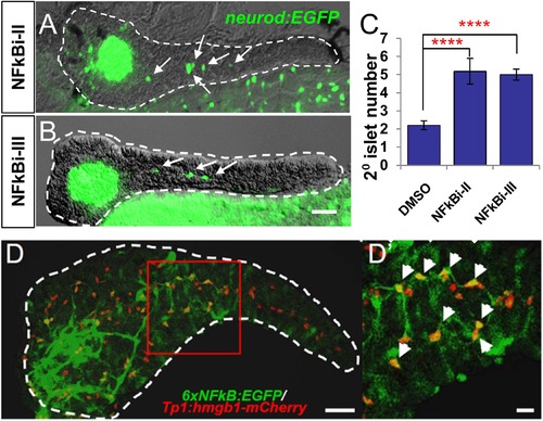
NF-κB pathway inhibition induces endocrine differentiation. (A, B) Representative confocal images - brightfield and fluorescence images merged - of dissected pancreata (dashed lines) from neurod:EGFP transgenic larvae treated from 3 to 5 dpf with NF-κB signaling inhibitor II (A) or III (B). Both inhibitors induced precocious secondary islet formation (white arrows). (C) Secondary islet numbers were quantified and plotted relative to vehicle control (0.1% DMSO). n = 5-10 larvae per condition, experiment was repeated 3 times. Error bar, standard error. All p-values were calculated using Dunnett′s test. ****p < 0.0001. (D, D′) Representative in vivo confocal z-projection of pancreas (dashed lines) in 6xNFκB:EGFP ;Tp1:hmgb1:mCherry double transgenic larvae at 5 dpf showing co-labeling of the NF-κB reporter (green) and Notch reporter (red) in endocrine progenitor cells (arrows in D′), suggesting endocrine progenitors respond to both Notch and NF-κB signaling. Scale bars, 25 µm (D), 10 µm (D′). |
| Genes: | |
|---|---|
| Fish: | |
| Anatomical Term: | |
| Stage: | Day 5 |