
Fig. 8
- ID
- ZDB-FIG-151008-22
- Publication
- Hu et al., 2015 - Loss of DDB1 Leads to Transcriptional p53 Pathway Activation in Proliferating Cells, Cell Cycle Deregulation, and Apoptosis in Zebrafish Embryos
- Other Figures
- All Figure Page
- Back to All Figure Page
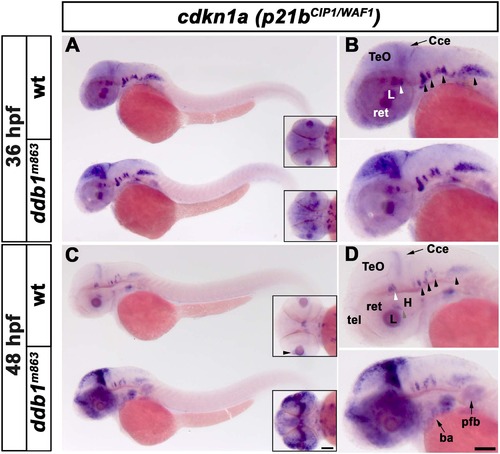
Increased p21bCIP1/WAF1 expression in homozygous ddb1m863 mutants. (A-B) Enhanced expression of p21bCIP1/WAF1 (cdkn1a) in ddb1m863 homozygous embryos in the proliferation regions of the optic tectum, compared to wild type siblings at 36 hpf. (C-D) At 48 hpf, p21bCIP1/WAF1 expression was strongly elevated in proliferation regions of the optic tectum, cerebellum, telencephalon, retina, branchial arches and pectoral fin bud in ddb1m863 mutants compared to wild type siblings. In contrast, expression of p21bCIP1/WAF1 in ventral hindbrain nuclei (arrow heads in B, D) was unaltered in ddb1m863 mutants. All views are lateral except for inserts (dorsal views). Abbreviations used: Cce, cerebellum; H, hypothalamus; L, lens; pfb, pectal fin bud; ret, retina; tel, telencephalon; TeO, tectum opticum. Anterior towards the left. Scale bar: 100 µm. |
| Gene: | |
|---|---|
| Fish: | |
| Anatomical Terms: | |
| Stage Range: | Prim-25 to Long-pec |